Summary
Physical Description
Ecology
Life History & Behaviour
Anatomy & Physiology
Evolution & Systematics
Biogeographic Distribution
Conservation & Threats
References & Links | 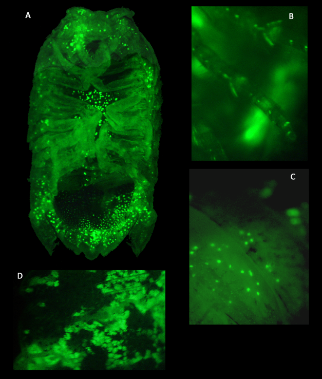 Anatomy & Physiology Anatomy & Physiology
As seen below, the sagital section of C. pelsarti provides insight into the anatomy of Sphaeromatid isopods. This is a hematoxylin and eosin stain which colours the nuclei of cells blue and other material pink. The section height shows the significant dorso-ventral flattening observed in this bilateral invertebrate. Unfortunately, due to the dense mineralisation of the outer cuticular layer, a clean section was difficult to obtain without smearing. The image below is a composite of eight separate images to build up a body length view due to poorer quality at high magnification.
 Sagital H&E section of Cymodoce cf. pelsarti labeled with identifiable features.
Chromatophores
Chromatophores are cells located below the epidermis and contain intracellular pigment granules and branched cytoplasmic processes (Ruppert et al. 2004). Many crustacea possess chromatophores which provide them some level of control over physical appearance. Chromatophores may change the appearance of an invdividual by either increasing in number or changing in size. The image located in the Physical Description section highlights the differences in appearance than can occur over a period of hours in individual isopods of the same species.
Chromatophores may form clumps of communicating cells called chromatophore organs. Within these clumps of cells, pigments may move around to particular areas to change the appearance of the whole group. The most effective colour change mechanism for chromatophore organs is for the pigments to spread out across the whole organ (darker) or become concentrated at the centre (lighter). The brown and black pigments observed in the chromatophores of C. pelsarti are hypothesised to be melanin, however multiple pigments may contribute to the colour of a single chromatophore. The control of these pigments is obtained through hormones released from the sinus gland (Ruppert et al. 2004).
Further investigation into the presence, pattern and structure of these chromatophores led to the testing of fluorescent response under UV excitation. Although it is known that many marine animals can express and detect fluorescent proteins presumably for communication, this feature has not yet been documented in the Sphaeromatid isopods. The collection of images below illustrate the bright green response to UV excitation from a desiccated specimen. It was observed that chromatophores were present in all areas of C. pelsarti except the pleopods and a higher concentration of chromatophores with defined pattern was present on the dorsal carapace. This is an exciting find as perhaps species may be more readily identified by chromatophore pattern or this may contribute to further understanding the ecology and environmental interactions of Cymodoce pelsarti.
Figure A-D: Green fluorescent response of Cymodoce pelsarti to UV excitation. Bright green spots are chromatophores.
Figure A: Three image composite providing a full ventral view.
Figure B: Presence of chromatophores in pereopods.
Figure C: Close-up of chromatophores on dorsal carapace.
Figure D: Close-up of chromatophores revealing chromatophore organs in which the fluorescing pigment is dispersed. |
|